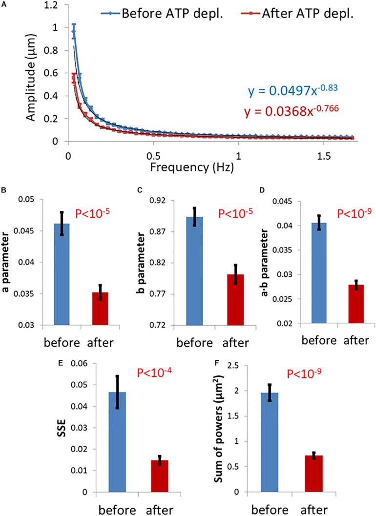
FIGURE 3.
Differential Interference Contrast (DIC) microscopy results of intracellular particles motion in live cells before and after adenosine triphosphate (ATP) depletion analyzed by amplitude spectral density (ASD) and power spectral density (PSD). (A) ASD analysis results in (n = 29) cells before (blue line) and after (red line) ATP depletion and the corresponding power fit parameters. (B–E) Power fit parameters: a, b, a⋅b, and SSE for the ASD analysis results in the cells [in (A)] before and after ATP depletion. (F) Power fit parameter “Sum of powers” for the PSD analysis results in the cells [in (A)] before and after ATP depletion. Error bars in panels (A–F) are SEM.

