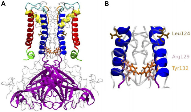
FIGURE 1.
Anatomy of KirBac3.1 S129R from the modeled KirBac3.1 S129R (A) The transmembrane portion of each monomer of KirBac3.1 is composed of four helices: slide helix (green), transmembrane helix 1 (red), pore helix (yellow), and transmembrane helix 2 (blue). The mutation S129R is situated at the level of the helix bundle (pink), at the bottom of the inner helix (B) Leu124 and Tyr132 are shown in color brown and orange respectively. Arg129 is shown in pink.

