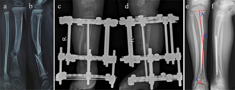
Fig. 3.
Preoperative frontal (a) and lateral (b) radiographs of an 11-month-old girl with Crawford type IV congenital pseudarthrosis of the right tibia with an intact fibula and associated neurofibromatosis type 1. Anteroposterior (c) and lateral (d) radiographs of the same patient presented at 1 week after combined surgery. Anteroposterior (e) and lateral (f) radiographs show the healed tibial pseudarthrosis with a normal fibula length and a normal medial proximal tibial angle (A, 87º), tibial diaphyseal valgus deformity (B, 10º), and lateral distal tibial angle (C, 85º) at 7 years after the combined surgery; the distal fibular physis was located at the level of the talar dome.

