
Figure 4:
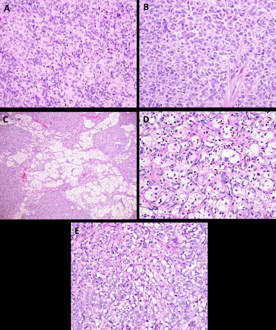
Cytoplasmic features in UTROSCT. A. Most have a small to moderate amount of pale, eosinophilic cytoplasm as seen in case 14 (H&E, 400x); B. Rhabdoid morphology in case 13 (H&E, 400x); C and D. Vacuolated/lipidized cytoplasm (C. case 2, H&E, 100x; D. case 12, H&E, 400x); E. Signet ring morphology in a single case (case 9, H&E, 400x).