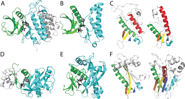
Figure 4.
Plasticity of distantly related protein kinase homologs. (A) Cki1 (PDB: 1csn) with bound Mg-ATP (gray sphere-black stick) in the catalytic cleft between the N-lobe (green, CATH 3.30.200) and the conserved core of the C-lobe (cyan, CATH 1.10.510) with additional elaborated helices (white). (B) Minimal kinase domain from OspG (PDB: 4q5hA) bound to MG-AMPPNP is colored as above. (C) Core protein kinase C-lobe SSEs (colored in rainbow from the N-terminus to the C-terminus) are common to Cki1 (left) and OspG (right). (D) ATP-grasp GART (PDB: 1kjqB; CATH 3.30.1 and 3.30.470) in complex with Mg-ADP is colored by the subdomain. An insertion in the N-lobe (white) replaces the N-terminal strands in protein kinases. The C-subdomain includes a pronounced β-sheet with similar topology to (E) SAICAR (PDB: 2gqrA, CATH 3.30.200 and 3.30.470) bound to Mg-ADP. (F) Common C-subdomain from SAICAR (rainbow) lacks the C-terminal helix (left), but a related SAICAR Itpkb includes the C-helix (PDB: 2aqx).

