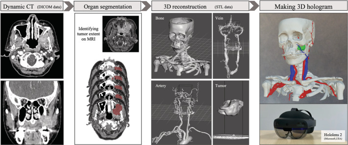
FIGURE 1.

Development of case specific 3D hologram. After organ segmentation of images from dynamic CT with MRI images as reference, 3D polygon files were exported and converted to 3D holograms for the see‐through head mount displays. Hololens 2 (Microsoft Corporation, Washington) was used as see‐through head mount displays in this study. 3D: three‐dimensional, CT: computed tomography, MRI: magnetic resonance imaging
