Figure 1.
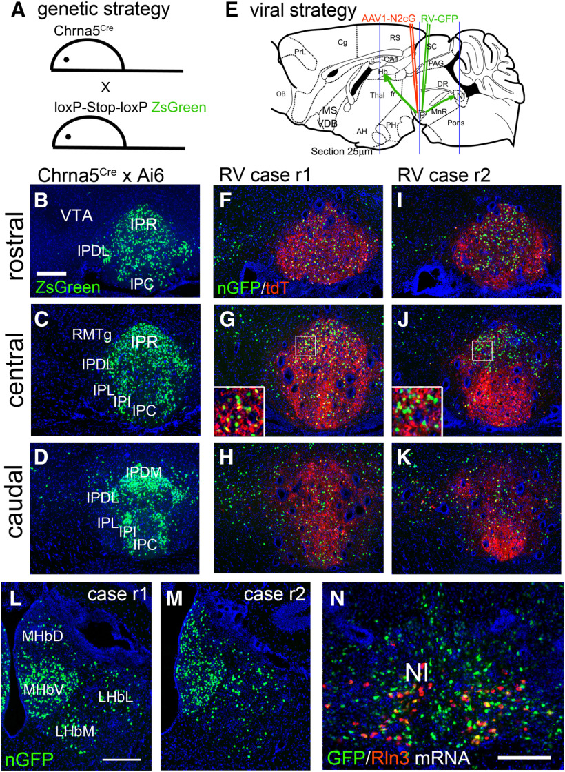
Genetic and viral strategy for transsynaptic labeling of IP afferents. A, A genetic strategy for labeling Cre-expressing neurons in the IP of Chrna5Cre mice. Chrna5Cre mice were interbred with the mouse strain Ai6, which conditionally expresses the fluorophore ZsGreen (Materials and Methods). B–D, ZsGreen expression in the rostral, central, and caudal IP of Chrna5Cre, Ai6 compound heterozygous mice, generated as shown in A. E, RV transsynaptic labeling strategy. A helper virus AAV1-N2cG was injected into the IP of Chrna5Cre mice, followed by RV three weeks later (Materials and Methods). The habenula and IP were examined for the expression of nuclear GFP (nGFP) expressed by RV. Blue lines indicate the planes of section for the habenula, IP, and NI (rostral to caudal). F–K, Imaging of nGFP expressed by RV and cytoplasmic tdTomato (tdT) expressed by the AAV helper virus in the rostral (F, I), central (G, J), and caudal (H, K) IP of two injected cases, r1 and r2 (retrograde 1 and 2). Insets in G, J show higher magnification of the boxed area. L, M, Expression of RV nGFP transsynaptic label in the habenula. Coronal sections correspond to bregma −1.7 in a standard atlas (Paxinos and Franklin, 2001). N, Dual-label FISH for RV-expressed GFP mRNA and Rln3 mRNA in the NI of injected Case r1. IP, interpeduncular nucleus; IPC, central part; IPDL, dorsolateral part; IPDM, dorsomedial part; IPI, intermediate part; IPL, lateral part; IPR, rostral part; LHbL, lateral habenula, lateral part; LHbM, lateral habenula, medial part; MHbD, medial habenula, dorsal part; MHbV, medial habenula, ventral part; NI, nucleus incertus; RMTg, rostromedial tegmental nucleus; VTA, ventral tegmental area. Scale bar: 200 μm (B, L, N).

