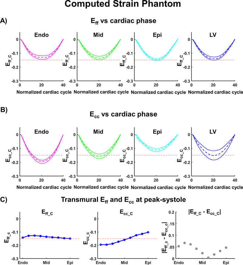
Figure 4:
Cardiac strain values for the computational phantom calculated after combining cDTI and DENSE. (A) Computed Myofiber strain (Eff_C) and (B) circumferential strain (Ecc_C) at endo, mid, epi layers, and across the left ventricular (LV) wall. The red dashed line provides a −0.15 strain reference; the colored dashed and dotted lines represent the median strain and the first and third quartiles across the corresponding layer. (C) Transmural distribution of Eff_C, Ecc_C, and the absolute difference between Eff_C and Ecc_C at peak systole. Blue solid lines represent the transmural median.

