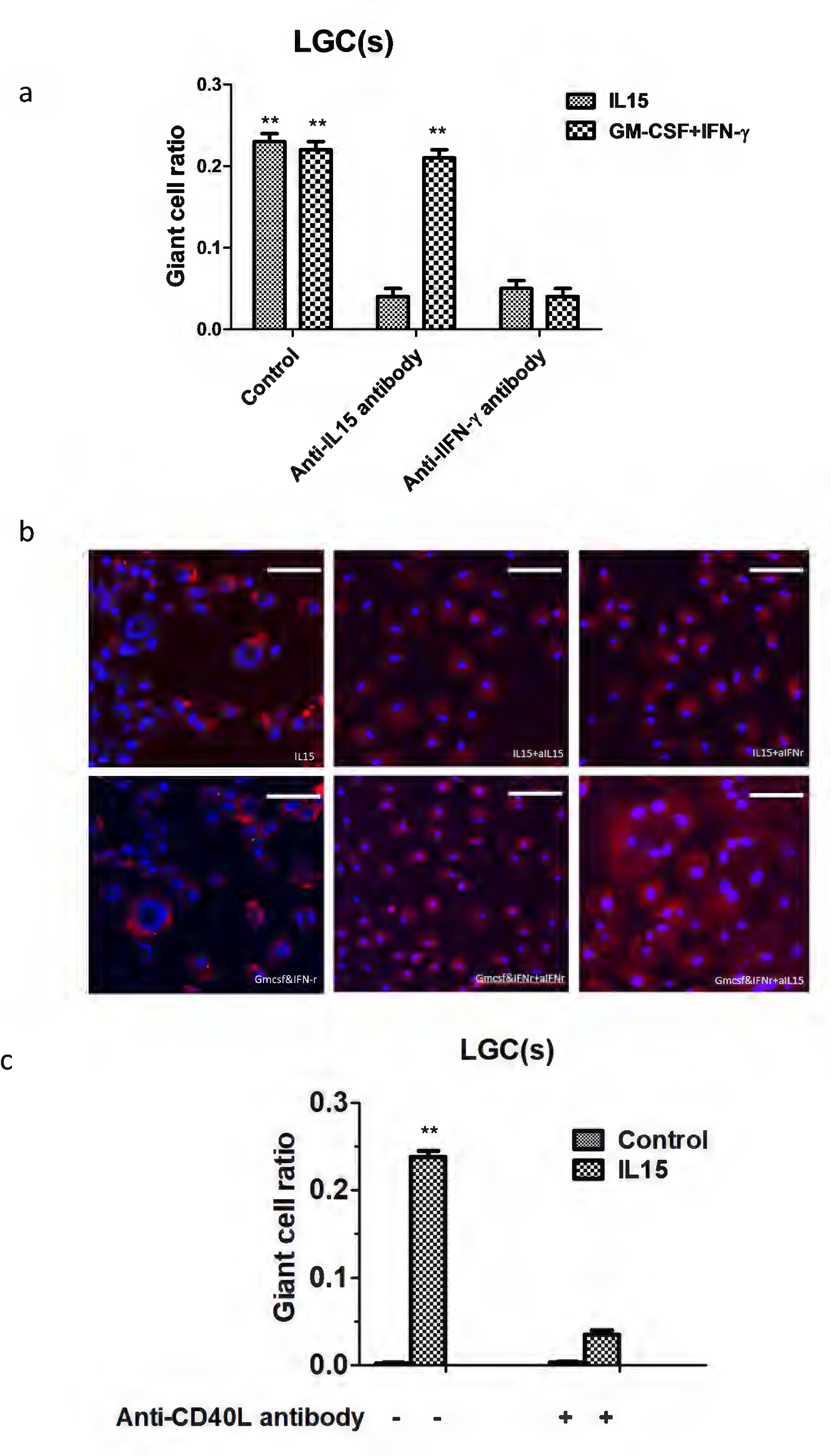
Figure 6.

The CD40-CD40L axis and IFN-γ were required for LGC formation. Highly purified monocytes isolated from T cell-depleted PBMC were cultured with the indicated concentration of rhIL15. The indicated antibodies (10 ug/ml) were added to the culture medium (a, b), in addition to exogenous sCD40L (3 ug/ml) (c). Values represent the mean giant cell ratio, and error bars indicate the standard mean of the error (n = 3 independent cultures). Scale bar: 100μm
