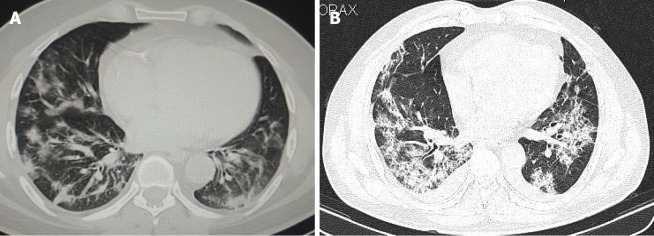
Figure 2.
Axial high-resolution computed tomography images of chest. A: Axial high-resolution computed tomography (CT) image of chest on day 5 after symptom onset demonstrating peripheral predominant consolidation pattern with areas of ground glass opacification in bilateral lower lobes in a patient with coronavirus disease 2019 pneumonia; B: Axial high-resolution CT image of chest on day 9 after symptom onset demonstrating extensive consolidation predominantly in basal segments of bilateral lower lobes in a patient with coronavirus disease 2019 related pulmonary syndrome. Note the bilateral pleural effusions which is an atypical finding in coronavirus disease 2019.

