
Figure 1. Determining basal CaMKII isoform expression in human and murine endothelium.
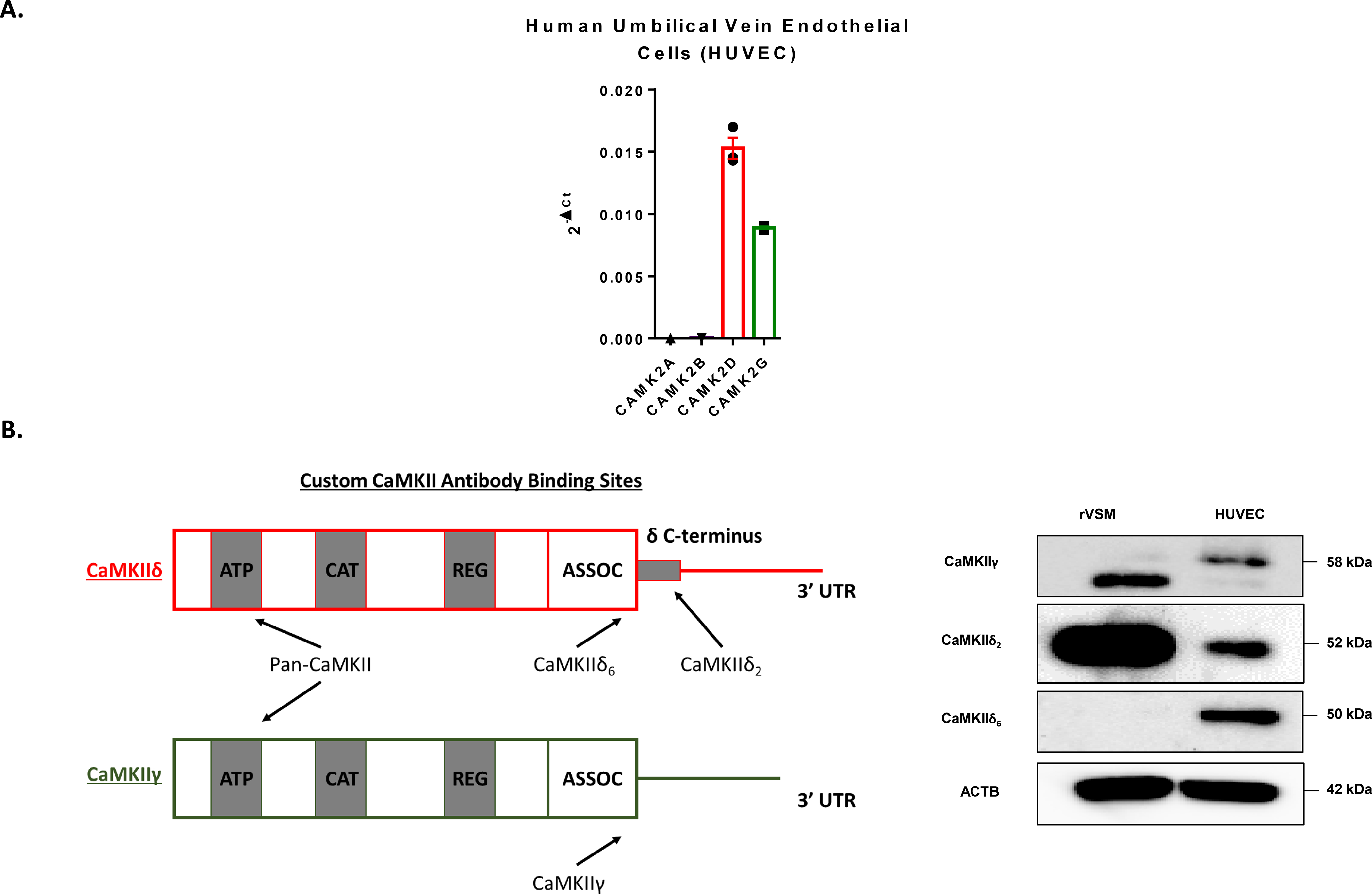
A. qPCR analysis of CaMKII subtypes CAMK2A, CAMK2B, CAMK2D, and CAMK2G in cultured HUVEC normalized to GAPDH. n = 3. B. Diagram of custom CaMKII antibody binding sites on CaMKIIδ and γ recognizing unique splice isoforms. Immunoblot for the variable domain of γ and the C-terminal tail of CaMKIIδ reveals differential isoform expression in endothelial cells when compared to rat vascular smooth muscle cells (rVSM). n = 3.