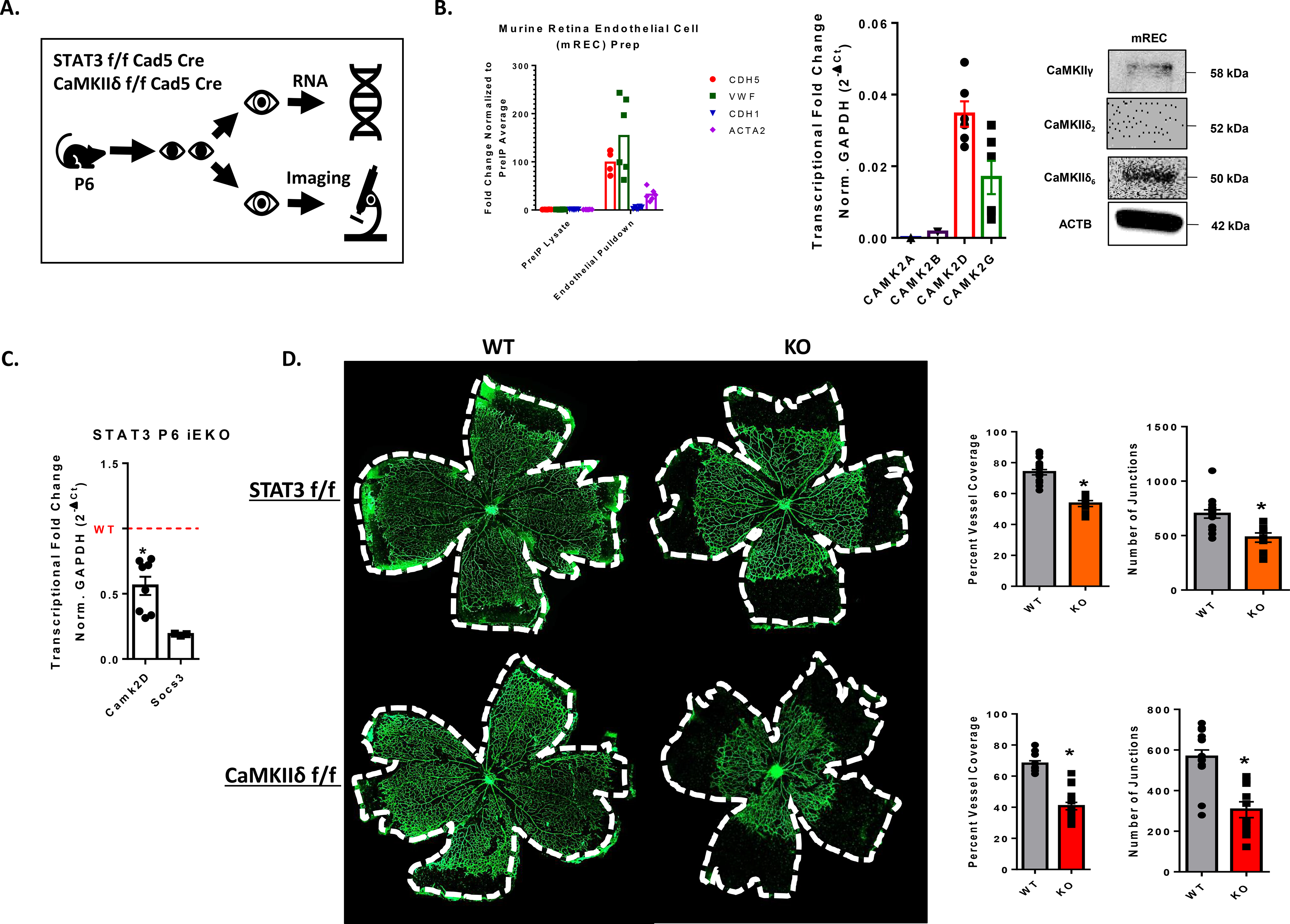
Figure 7. STAT3-dependent expression of CaMKIIδ promotes angiogenesis in vivo.

A. Schematic for strategy for measuring in vivo angiogenesis. Pups were oral gavaged with10 μL of 100 mg/mL tamoxifen at postnatal day 1, 2, and 3. At postnatal day 6, mice eyes were either collected and fixed for whole mount imaging or digested with collagenase solution and incubated with anti-CD31 beads for RNA isolation. B. qPCR analysis of endothelial marker genes (Cdh5, Vwf), epithelial marker Cdh1, and pericyte/VSM marker Acta2 expression in RNA isolated from antiCD31-pulldown lysates and non-bound fraction lysates extracted from murine retinas at postnatal day 6 (P6). qPCR analysis of Camk2a, Camk2b, Camk2d, and Camk2g expression in isolated murine retinal endothelial cells (mREC) normalized to GAPDH, n = 3. Western blot analysis of antiCD31-pulldown lysates with custom CaMKII antibodies targeted to CaMKIIδ2, CaMKIIδ6, and CaMKIIγ isoforms. n = 3. C. RNA from CD31-pulldown enriched retinal endothelial cells was used for qPCR analysis to detect relative amounts of CAMK2D in our inducible STAT3 loss of function animals. Data normalized to GAPDH transcript levels and Socs3 was utilized as a positive control for loss of STAT3 function. n = 3, Student’s t-test, (*) p< 0.05. D. Whole mount retinas from P6 endothelial-specific STAT3 loss of function mice and CaMKIIδ knockout mice. Retina vessels lableled with AlexaFluor488-IsolectinB4 and imaged using 4x tiling with the Cytation5 plate reader and imager. Percent Vessel coverage was calculated by normalizing the area covered by vessels to the area of the total retinal bed. Number of junctions was quantified using Angiotool2.0 software. n = 3 litters each, Student’s t-test, (*) p<0.05.
