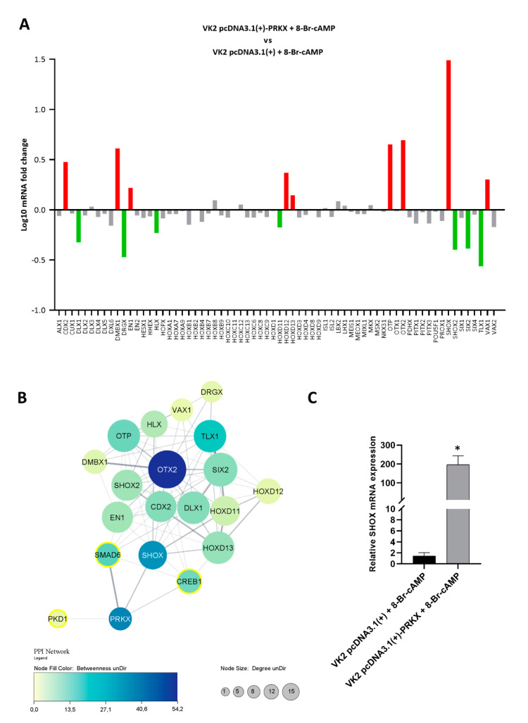
Figure 6.
PRKX overexpression influences the expression of HOX genes in VK2 E6/E7 cells. (A) Recapitulating graph of RT-qPCR screening for HOX gene mRNA level alterations in VK2 E6/E7 cells transfected with pcDNA3.1(+)-PRKX after 8-Br-cAMP treatment. Relative fold changes represented on a log10 scale are referred to as VK2 E6/E7 cells transfected with empty vector as the baseline. Gene expression variation was considered significant for fold changes ≤0.6 and ≥1.3. Significantly upregulated and downregulated genes are indicated as red and green columns, respectively. (B) PPI network showing interactions between PRKX, its principal downstream substrates SMAD6, CREB1, PKD1, and HOX genes’ products that were deregulated from the RT-qPCR array analysis. Node size is directly proportional to connectivity degree and node colour is correlated to betweenness value, with a darker colour corresponding to a higher value. PRKX substrates are circled in yellow. (C) RT-qPCR analysis showing SHOX mRNA levels in VK2 E6/E7 transfected with PRKX expression vector (pcDNA3.1(+)-PRKX) or empty vector as control (pcDNA3.1(+)) and treated with 8-Br-cAMP (mean ± SD; n = 2; * p < 0.05).

