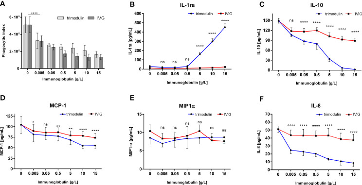
Figure 3.
Immune modulation in COVID-19-like model by IVIG and trimodulin preparation. (A) HL60 cells were incubated for 1 h with SARS-CoV-2 spike protein coated beads opsonized with COVID-19 plasma. IVIG (IgG Next Generation, Biotest AG) or trimodulin (Biotest AG) was added in the indicated concentrations to the cell immune complex mixture. Phagocytosis of SARS-CoV-2-like immune complex was measured with trimodulin (light gray bars) or IVIG (dark gray bars) addition. (B–F) Same as (A) instead phagocytosis cytokine release into cell culture supernatant was measured with trimodulin (dots, blue line) or IVIG (square, red line) addition. IL1-ra (B), IL-10 (C), MCP-1 (D), MIP-1α (E), and IL-8 (F) were measured. Values represent mean of six independent experiments. Statistics: Two way ANOVA; Tukey’s multiple comparisons test, 95% confidence interval. ns, not significant, *p ≤ 0.05, **p ≤ 0.01, ****p ≤ 0.0001.

