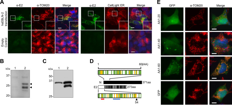
FIG 2.
Mitochondrial targeting of the E2 protein. (A) Subcellular localization of E2. OL cells were transfected with the E2 expression plasmid or empty plasmid and stained with an anti-E2 antibody. An anti-TOM20 antibody and CellLight endoplasmic reticulum (ER)-red fluorescent protein (RFP) were used to visualize mitochondria and the ER, respectively. Cell nuclei were stained with 4′,6-diamidino-2-phenylindole (DAPI) and observed by fluorescence microscopy. Bars, 10 μm. (B and C) The E2 protein is cleaved in transfected cells. (B) 293T cells were transfected with the control (lane 1) or E2 expression plasmid (lane 2), and the E2 protein was detected by Western blotting with an anti-E2 antibody. (C) 293T cells were transfected with the E2 expression plasmid tagged with Flag at the C (lane 1) or N terminus (lane 2) of the protein, and the E2 protein was detected by Western blotting with an anti-Flag antibody. (D) The E2 protein contains a mitochondrial targeting signal. The full-length and N-terminal 60 amino residues of the Borna disease virus 1 (BoDV-1) N and E2 proteins are shown in the schematic diagram. Sites in the E2 protein homologous or similar to those in the N protein are indicated by black lines. The green line indicates hydrophobic amino acids, and the yellow line indicates basic amino acids. The arrowheads indicate the predicted cleavage positions by mitochondrial processing peptidase predicted by both Mitoprot and MitoFates. The red and blue lines indicate the binding site of TOM20 and the positively charged amphiphilicity high score region predicted by MitoFates, respectively. N, BoDV-1 N; E2, hsEBLN-2. (E) The N terminus of BoDV-1 N contains a putative mitochondrial targeting signal (MTS). OL cells were transfected with plasmids expressing green fluorescent protein (GFP) fused with the N-terminal 20 amino acids (aa) (AA1-20), 40 aa (AA1-40), and 60 aa (AA1-60), and their localization was detected by fluorescence microscopy after staining with an anti-TOM20 antibody. Bars, 10 μm.

