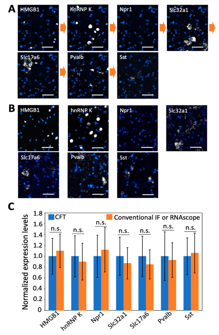
Figure 7.
(A) Proteins HMGB1 and hnRNP K together with mRNA Npr 1, Slc32a1, Slc17a6, Pvalb, and Sst are stained sequentially with tyramide-N3-Cy5 in a fixed frozen mouse spinal cord tissue. (B) The same two proteins and five mRNA are stained in seven different fixed frozen mouse spinal cord tissues by conventional immunofluorescence (IF) or RNAscope. (C) Comparison of the normalized single-cell expression levels obtained in (A,B) (n = 30 cells). Error bars generated using standard error of the mean. n. s., p > 0.4. Scale bar, 40 µm.

