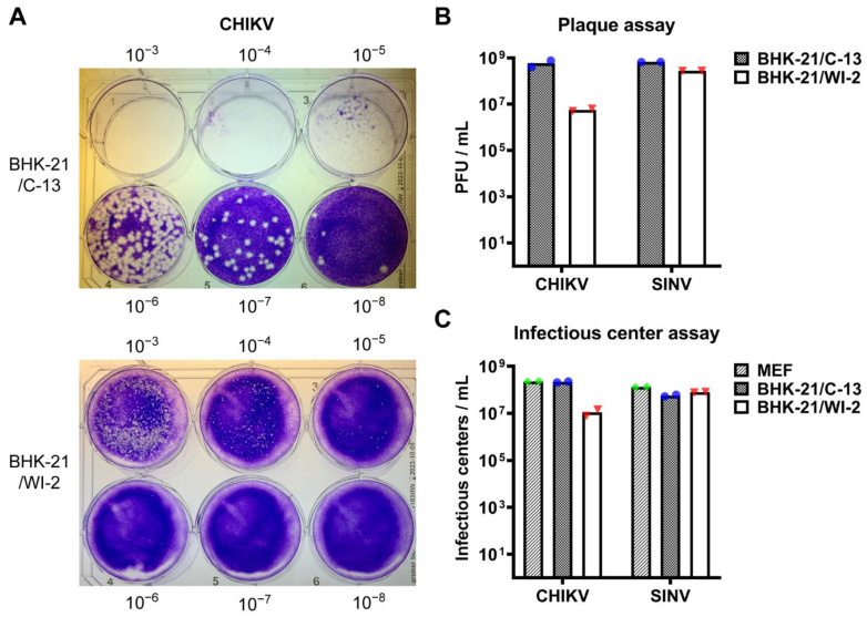
Figure 1.
Infectivity of CHIKV in BHK-21/C-13 and BHK-21/WI-2 cells. (A,B) BHK-21/C-13 cells or BHK-21/WI-2 cells were infected in parallel with serial dilutions of CHIKV or SINV and overlaid with medium containing 0.5% CMC. Plaques were fixed and stained at 48 h post-infection. Representative images from one of 2 independent experiments are shown in (A). The bar graph in (B) shows the mean and individual data points of 2 independent experiments. (C) Infectious center assay. MEF, BHK-21/C-13, or BHK-21/WI-2 cells were infected in parallel with serial dilutions of CHIKV or SINV. Primary infection was quantitated by immunofluorescence. Data shown are the mean and individual data points of 2 independent experiments.

