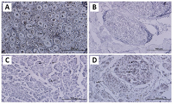
Figure 3.

Immunohistochemistry staining of anti-NF-200.
Sections of native (A) and decellularized nerve (B) were stained by the anti-NF-200 antibody. Immunohistochemistry showed that nerve fibers were removed by the decellularization process (B). After 8 weeks, staining of NA (C) and ANX (D) grafts showed that many NF-200 positive myelinated axons were observed throughout the grafts, and in the xenografts as well. Arrows indicate the axons. Scale bars: 100 μm. ANX: Acellular nerve xenografts; NA: nerve autograft; NF-200: neurofilament 200.
