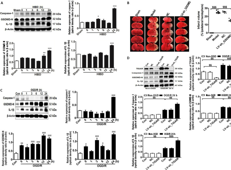
Figure 2.
TIGAR knockdown increases the activation of pyroptosis in the neonatal cortex and HAPI microglial cells of HIBD models in vivo and in vitro.
(A) Quantification of caspase-1, GSDMD-N, and IL-1β in neonatal rat cortex 24 hours after HIBD. (B) The lesion infarct was detected using 2,3,5-triphenyltetrazolium chloride staining 24 hours after HIBD. Red indicates normal tissue and white indicates ischemic tissue. (C) Quantification of caspase-1, GSDMD-N, and IL-1β in HAPI microglial cells 24 hours after OGD/R. (D) Quantification of TIGAR, caspase-1, GSDMD-N, and IL-1β in HAPI microglial cells with or without LV-sh_TIGAR treatment 24 hours after OGD/R. Data are expressed as the mean ± SEM (A, C, D: n = 3 animals/group; B: n = 6 animals/group). *P < 0.05, **P < 0.01, ***P < 0.001, vs. sham group; #P < 0.05, ##P < 0.01, ###P < 0.001, vs. control group; §§P < 0.01, §§§P < 0.001 (one-way analysis of variance followed by Bonferroni’s multiple-comparisons post hoc test). Con: Control; GSDMD-N: gasdermin D N-terminal; HAPI: highly aggressively proliferating immortalized; HIBD: hypoxic-ischemic brain damage; IL-1β: interleukin-1β; LV-sh_TIGAR: vector containing short hairpin RNA of TIGAR; Model: HIBD; NC: LV-sh-scramble; OGD(/R): oxygen and glucose deprivation(/reoxygenation); TIGAR: TP53 induced glycolysis and apoptosis regulator.

