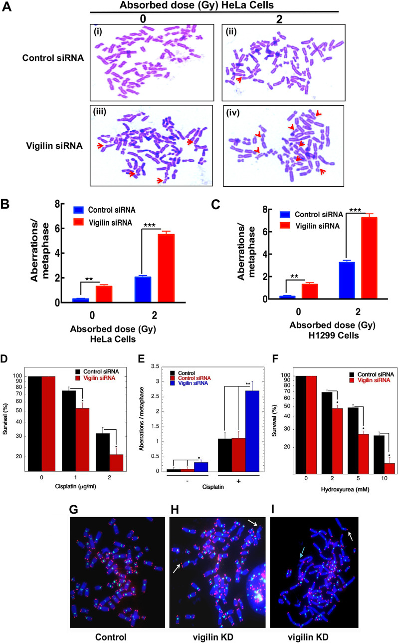
FIG 2.
Chromosome aberration analysis. (A) Representative images of metaphase spread of cells in control and vigilin knockdown HeLa cells showing increased spontaneous and IR-induced metaphase chromosome aberrations. (B and C) Quantification of chromosome aberrations seen in HeLa and H1299 cells, respectively, with and without depletion of vigilin. Quantification represents aberrations from 30 metaphase spreads of three independent experiments. (D to F) Clonogenic cell survival assay and metaphase chromosome aberrations of vigilin-depleted HeLa cells treated with the indicated concentrations of cisplatin and hydroxyurea. Vigilin-depleted cells show decreased survival and increased chromosome aberrations upon treatment with cisplatin and hydroxyurea compared to control cells. The error bars represent the standard deviations from three independent experiments. (G) Human metaphase chromosomes showing telomeres in red and centromeres in green detected by FISH using specific probes in control and vigilin knockdown cells. (H and I) Metaphase chromosomes from vigilin-depleted cells showing dicentrics indicated by white arrows and loss of telomere signals indicated by green arrow.

