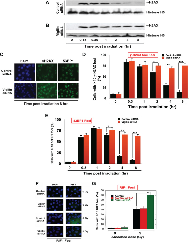
FIG 4.
Depletion of vigilin alters the DNA damage response. (A and B) HeLa cells were transfected with either control siRNA or vigilin siRNA followed by irradiation with 2 Gy and then analyzed by Western blotting for the appearance/disappearance of γ-H2AX after release at different time points. H3 was used as a loading control. (C) Control and vigilin-depleted cells were irradiated with 4 Gy, fixed after IR treatment, and stained for γ-H2AX and 53BP1 foci. (D and E) Quantification of percentage of cells with γ-H2AX and 53BP1 foci in control and vigilin knockdown cells at different time points postirradiation. For each time point, 100 cells were analyzed, and the number of cells with more than 10 foci was plotted against time. (F and G) HeLa cells were irradiated with 5 Gy, and RIF1 foci were quantified for RIF1 foci in control and vigilin-depleted HeLa cells with and without IR treatment. Vigilin-depleted cells show higher levels of residual γ-H2AX, 53BP1, and RIF1 foci postirradiation compared to control cells. Error bars represent standard deviations from three independent experiments.

