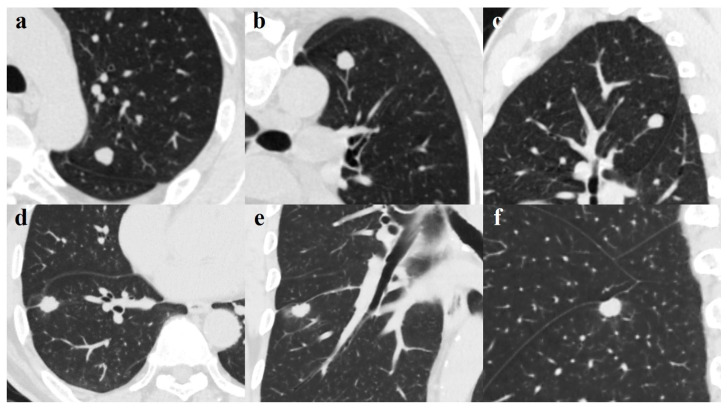
Figure 3.
(a–c). A 49-year-old man with pulmonary tuberculoma in the upper lobe of the left lung. Axial image (a) showing a round-like well-defined solid nodule measuring 1.14 cm; blood vessel convergency can be seen on the reconstructed coronal (b) and sagittal (c) views. The lesion was confirmed on pathological diagnosis as a tuberculoma. d-f. A 65-year-old man with lung adenocarcinoma in the right lower lobe. Axial image (d) demonstrates a well-defined juxta-fissural solid nodule measuring 1.47 cm with lobulated margin, short burr, and pleural indentation sign. Perilesional ground-glass opacification and a pleural indentation sign can be seen on the coronal (e) and sagittal (f) views. The lesion was proven on pathological diagnosis to be a moderately differentiated adenocarcinoma.

