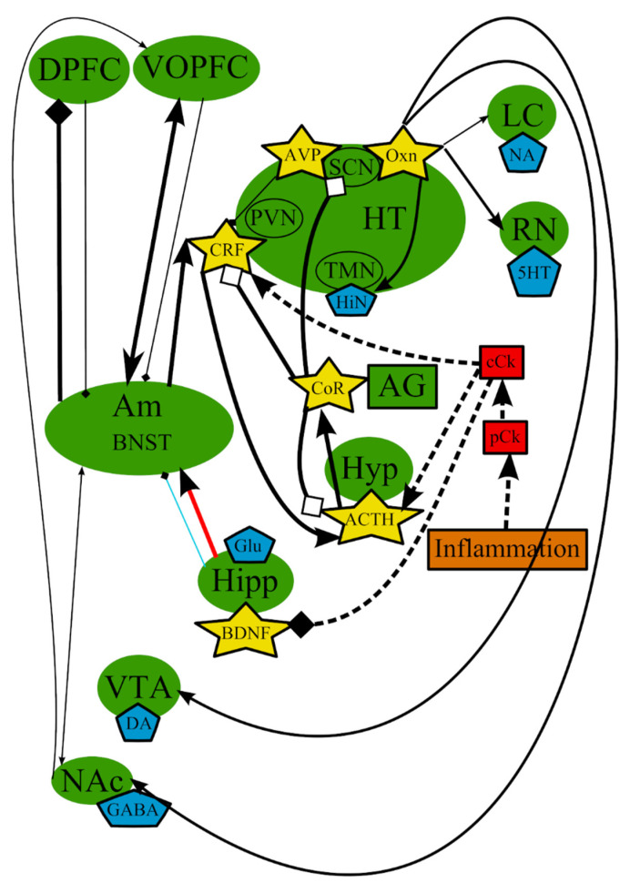
Figure 3.
The inflammation/cytokine hypothesis: main structures, neurotransmitters, biologically active substances, interactions, and external factors. ACTH—adrenocorticotropic hormone; AG—adrenal gland; Am—amygdala; AVP—arginine-vasopressin; BDNF—brain-derived neurotrophic factor; BNST—bed nuclei stria terminalis; cCk—central pro-inflammatory cytokines; CoR—cortisol; CRF—corticotropin-releasing factor; DA—dopamine; DPFC—dorsal prefrontal cortex; Glu—glutamine; GABA—gamma-aminobutyric acid; HiN—histamine; Hipp—hippocampus; HT—hypothalamus; 5-HT—serotonin; Hyp—hypophysis; LC—locus ceruleus; NA—noradrenaline; NAc—nucleus accumbens; Oxn—orexin; SCN—suprachiasmatic nucleus; TMN—tuberomammilar nucleus; RN—raphe nucleus; pCk—peripheral pro-inflammatory cytokines; PVN—paraventricular nucleus; VOPFC—ventral and orbital prefrontal cortex; VTA—ventral tegmental area; -> (arrow): activating effect; -<> (rhombus): a black rhombus—inhibitory effect; a white rhombus: an effect is blocked or ineffective because the receptor is not sensitive; thick line—effect is increased; thin line—effect is decreased; medium thickness line—effect is not changed or alterations of the effect are unknown; red line—noradrenaline effect (most of them were omitted to simplify the figure); blue line—serotonin effect (most of them were omitted to simplify the figure); black line—various neurotransmitters or neuropeptides; dotted lines—influence of external factors.

