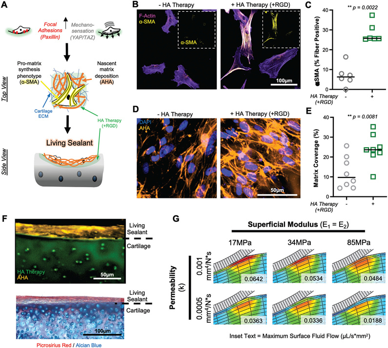
Figure 6.
HA therapy promotes MSC-guided matrix synthesis on degenerated cartilage tissue. A) Schematic depicting how adhesion and mechanosensation drive α-smooth muscle actin (α-SMA) positive stress fiber formation and deposition of nascent matrix, to form a living sealant on damaged cartilage. B) F-actin (magenta) and α-SMA (yellow) staining of MSCs cultured (t = 7 d) on degenerated cartilage with and without HA therapy (+RGD). Inset shows α-SMA channel alone. Scale bar = 100 μm. C) Percent of cells positive for α-SMA stress fibers. n = 6 replicates per group, n > 30 cells per group per replicate. D) Nascent matrix deposition, visualized by staining of azidohomoalanine (AHA; orange), on cartilage with and without HA therapy (+RGD) after 7 d of culture. Scale bar = 50 μm. E) Percent of cartilage area covered by nascent matrix. n = 7 replicates, n = 5 images per replicate. F) Top: Cross-sectional view of nascent matrix (orange) covering the surface of cartilage explant treated with HA therapy (green). Bottom: Histological image of cartilage explant with HA therapy, seeded with MSCs, and cultured for 7 d, showing the formation of a living barrier. Collagenous (picrosirius red) and glycosaminoglycan (Alcian blue) visualized. G) Finite element modeling of orthotropic sealant layer showing impact of sealant permeability and tensile modulus (E1 = E2, both parallel to articular surface) on fluid flux (μL/s*mm2) at the cartilage surface. Inset text represents maximum fluid flow at the cartilage surface (top 20 μm).

