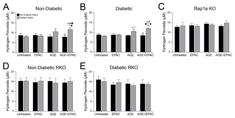
Figure 8.
AGE/RAGE signaling and Rap1a expression impacted hydrogen peroxide levels in cardiac fibroblasts. Cardiac fibroblasts isolated from (A) non-diabetic, (B) diabetic, (C) Rap1a KO, (D) non-diabetic RKO, and (E) diabetic RKO mouse hearts. Fibroblasts were embedded in non-diabetic (black bars) and diabetic (grey bars) matrices for 7 days and treated with EPAC (100 µM), exogenous AGEs (0.05 mg/mL) and EPAC+exogenous AGEs (100 µM and 0.05 mg/mL, respectively). Protein lysates were collected and assessed for hydrogen peroxide concentration. Mean ± SEM were graphed with n = 5–10 and statistical analysis consisted of a two-way ANOVA, followed by a Fisher’s Least Significant Difference post hoc. Symbols above each bar indicate significance between a specific group (p < 0.05; * vs. untreated in non-db, + vs. untreated in db, # vs. EPAC in non-db, ◆ vs. EPAC in db, ▽ vs. AGE in non-db, ⊗ vs. AGE in db, and • vs. AGE+EPAC in non-db). Untreated treatment groups embedded in non-diabetic collagen were initially presented in Figure 7.

