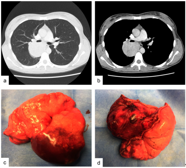
Figure 1.
Case 1. (a,b) contrast enhanced chest CT-scan showing an hypodense soft-tissue lesion in the size of mm 50 by 65 mm located between the posterior segment of the right upper lobe and lower lobe upper segment. (c,d) operative specimen: right upper lobe and lower lobe upper segment after surgical excision.

