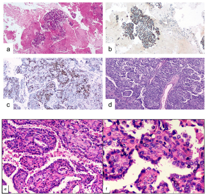
Figure 2.
Case 1, microscopic findings. (a) cyto-inclusion: spindle cells with focal atypia in the adenomorphic and papillary pattern (hematoxylin-eosin, 10×). (b) cyto-inclusion: positivity of the neoplastic cells for TTF-1 (10×). (c) round and spindle cells aggregated in papillae solid nests ex-pressed focal positivity for progesterone-receptor (10×). (d) histological features of PSP, with varied proportions of sclerotic, solid, papillary and hemorrhagic patterns (hematoxylin-eosin, 10×). (e) histological detail of the PSP architecture (hematoxylin-eosin, 20×). (f) histological detail of the PSP architecture (hematoxylin-eosin, 40×).

