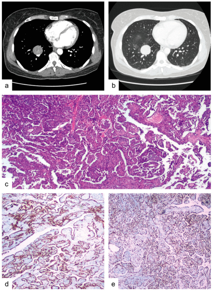
Figure 3.
Case 2. (a) contrast enhanced chest CT-scan revealing a 35 mm in diameter oval-shaped solid mass with well-defined borders in the right lower lobe, medial basal segment. (b) the above-mentioned mass with surrounding ground-glass opacities. (c) histological features of PSP, including both cell types (epithelial and stromal) aggregated mainly in a papillary/sclerotic pattern (85%) with a smaller solid pattern component (15%), (hematoxylin-eosin, 10×). (d) positivity of the neo-plastic cells for CK-7 (10×). (e) positivity of the majority of the spindle cells for TTF-1 (10×).

