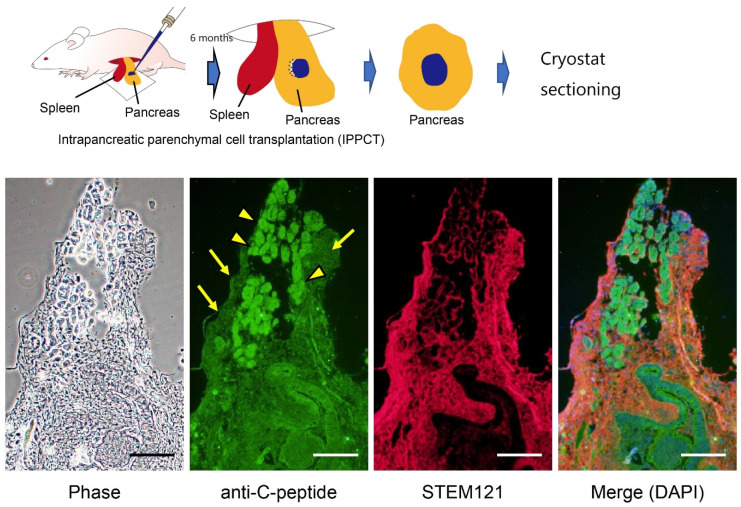
Figure 5.
Production of insulin in the grafts after intrapancreatic parenchymal cell transplantation in nude mice. In vitro cultured cell masses (on day 15 after induction; see Figure 4) derived from HDDPC-NSCs were transplanted into the pancreas of nude mice under a dissecting microscope using a glass micropipette (shown above). Six months after grafting, the pancreas was dissected and subjected to cryostat sectioning. Immunostaining using antibodies against insulin (C-peptide) revealed the presence of insulin-positive cells (arrowed) in the graft. Immunostaining using antibodies against STEM121, human cell-specific antibodies, revealed the presence of HDDPC-NSC-derived cells. Arrowheads indicate mouse pancreatic islets. We obtained these images from experiments performed exclusively for this review. Scale = 100 μm.

