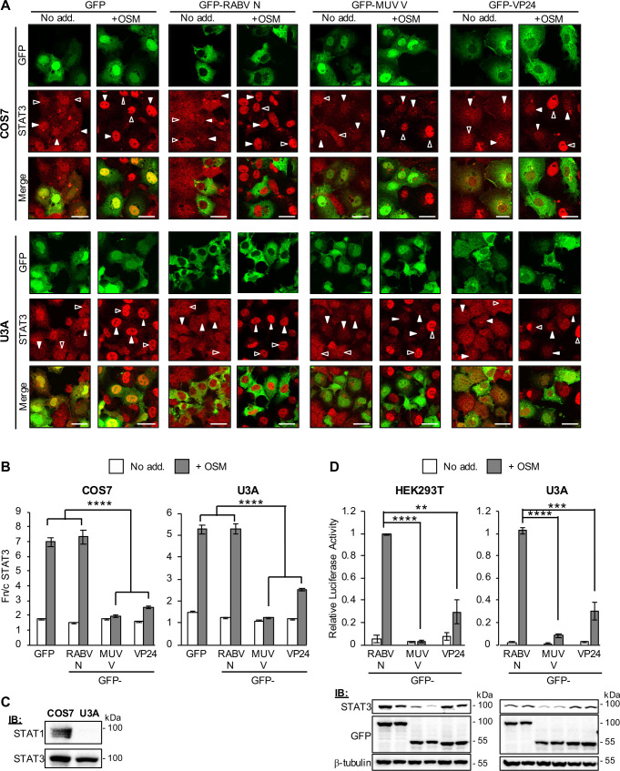
Fig 4. EBOV VP24 antagonises STAT3 independently of STAT1.
(A, B) COS7 (upper panel) or U3A (lower panel) cells transfected to express the indicated proteins were treated 24 h post-transfection with or without OSM (10 ng/ml, 15 min) before fixation, immunofluorescent staining for STAT3 (red) and CLSM (A) to determine the Fn/c for STAT3 (B; mean ± SEM, n ≥ 36 cells for each condition; results are from a single assay representative of two independent assays). Filled and unfilled arrowheads indicate cells with or without, respectively, detectable expression of the transfected protein. Scale bars, 30 μm. MUV V, Mumps virus V protein. (C) Lysates of COS7 and U3A cells were analysed by immunoblotting (IB) for STAT1 and STAT3. (D) upper panel: HEK293T or U3A cells co-transfected with m67-LUC and pRL-TK plasmids, and plasmids to express the indicated proteins, were treated 16 h post-transfection with or without OSM (10 ng/ml, 8 h) before determination of relative luciferase activity (mean ± SEM; n = 3 independent assays); lower panel: cell lysates used in a representative assay were analysed by IB using antibodies against the indicated proteins. Statistical analysis used Student’s t-test; **, p<0.01; ***, p < 0.001; ****, p < 0.0001.

