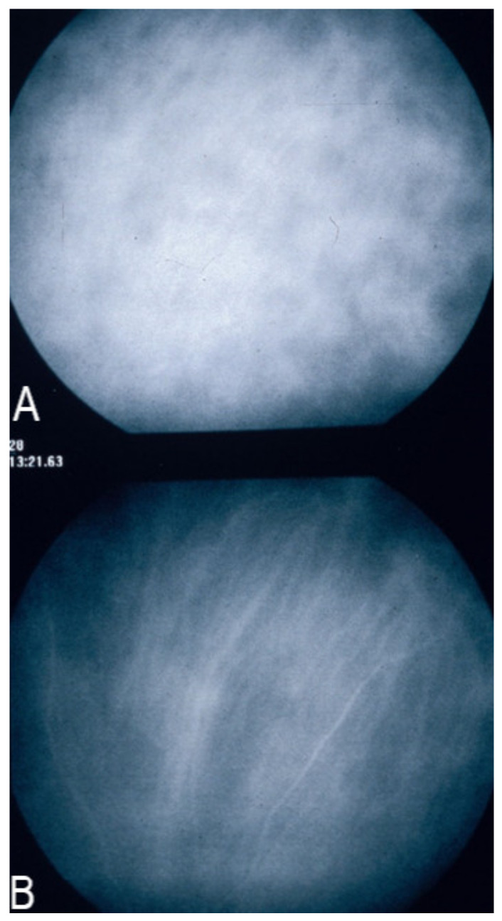
Figure 6.
Indocyanine green angiography (ICGA), type 2 pattern found in stromal choroiditis. (A): Typical round HDDs of similar size evenly distributed caused by the presence of inflammatory foci that impair the diffusion of the ICG dye showing granulomas in negative in case of Vogt-Koyanagi-Harada (VKH) disease. Choroidal vessels are no more distinct and appear fuzzy with diffuse hyperfluorescence hiding the HDDs. (B): After three days of intravenous corticosteroids, the choroidal vessels are again distinctly visible, and HDDs have partially resolved.

