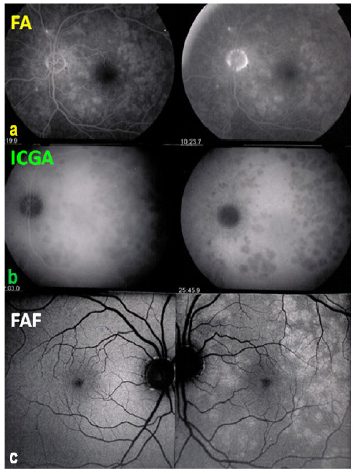
Figure 7.
Multimodal imaging of a case of MEWDS. FA (a) shows faint FA hyperfluorescence probably caused by outer retinal ischaemia. ICGA (b) shows extensive zones of hypofluorescence due to choriocapillaris non-perfusion. FAF (c) shows extensive BAF hyperautofluorescence that co-localise with ICGA hypofluorescent areas and is produced by the loss of the photopigment screen unmasking the normal underlying RPE autofluorescence.

