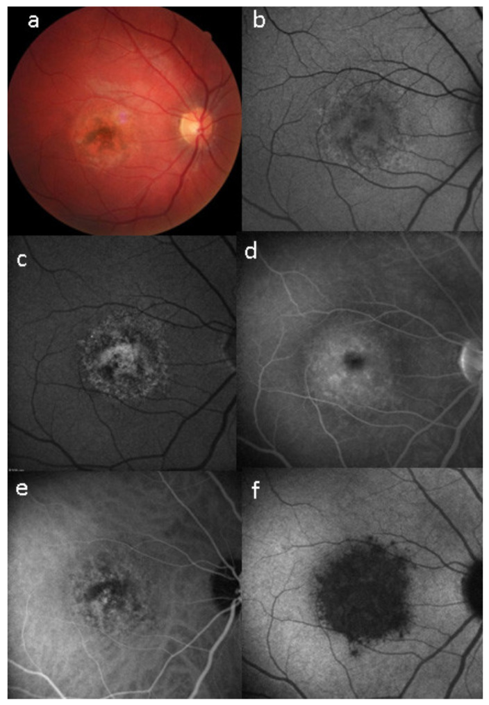
Figure 33.
Case of UIAM at presentation: (a) fundus photography showing a reddish foveal detachment surrounded with an irregular, circular area of greyish RPE discoloration. (b) BL-FAF shows a light hypoautofluorescence in the macular area. (c,d) FA early and late phases reveal late hyperfluorescence due to the leakage and pooling in the neurosensory foveal detachment. (e,f) ICGA early and late phases reveal persistent hypofluorescent area surrounded, in the late frame, by a hyperfluorescent ring, sign of hyperpermeability of the choroidal vessels.

