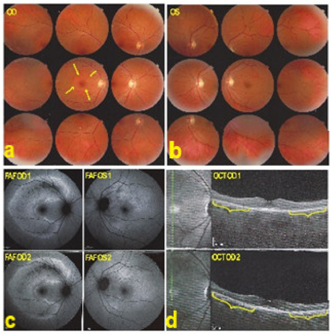
Figure 36.
AZOOR. Fundus photograph showing a pale ring around the fovea OD (arrows) (a) and normal OS (b). In (c) Fundus autofluorescence (FAF) shows an extensive C-shaped hyperautofluorescent area around the fovea with dots of hypoautofluoresce within this zone OD. (FAF OD 1) Left hyperautofluorescence parapapillary inferiorly and supero-temporally to the fovea. (FAF OS 1), corresponding to the loss of photoreceptors on SD OCT, extensive in OD (brackets in OCT OD 1 (d)), with a stable evolution after infliximab therapy (FAFOD/OS 2 and brackets in OCT OD 2 (d)).

