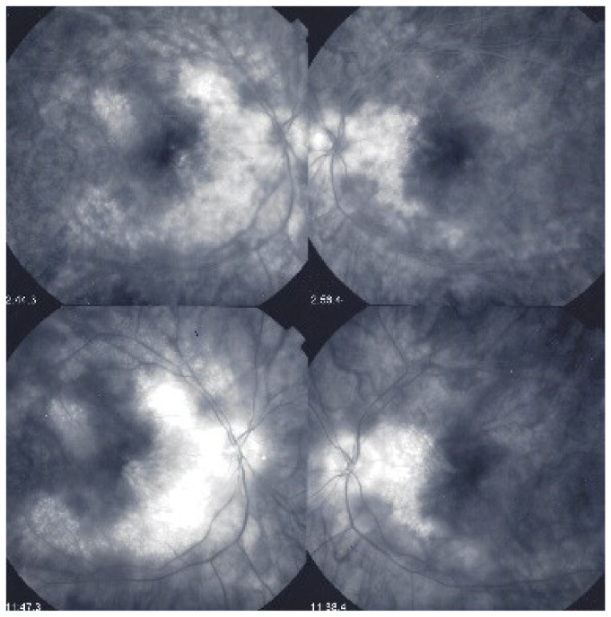
Figure 43.
Foveal sparing of diffuse retinal edema. This patient was followed for more than seven years for “neuroretinitis”, without treatment. There is massive retinal oedema that also involves the posterior pole, yet the fovea remains relatively spared, which explains the patient’s preserved visual acuity, while visual fields were severely impaired (Figure 44).

