Figure 1.
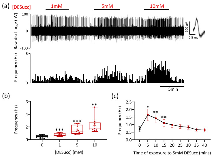
Succinate causes carotid body (CB) chemoafferent stimulation. (a) Example CB chemoafferent recording of the response to 1, 5, and 10 mM diethyl succinate (DESucc). Raw voltage is shown (upper) along with frequency histograms (lower). Overdrawn action potentials are inset, demonstrating single fibre discrimination. (b) Mean steady-state responses to DESucc at 1, 5, and 10 mM concentrations (n = 9 fibres, N = 4 animals). Data presented as box-whisker plots with median, mean (shown as +), the box representing the interquartile range and the whiskers extending to outliers. Single points represent individual fibres. ** and *** denote p < 0.01 and p < 0.001 vs. 0 mM, one-way repeated measures ANOVA with Dunnett’s post hoc test. (c) Time course of prolonged exposure to 5 mM DESucc (n = 11 fibres, N = 5 animals). Data presented as mean ± SEM. * and ** denote p < 0.05 and p < 0.01 vs. 0 min, one-way repeated measures ANOVA with Dunnett’s post hoc test.

