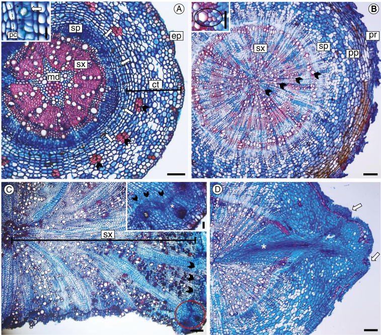
Fig. 2.
Root cross-sections at the secondary stage of development in Centaurea jacea (A), Euphorbia esula (B, D) and Knautia arvensis (C). The anatomical predictors used in the statistical analysis are shown. (A) Species with epidermis (ep) and cortex (ct) preserved; sclerified cortical cells (arrowheads) are scattered on the cortical parenchyma, and the endoderm (* inset) associated with secretory structures (arrows) constitutes the inner layer of the cortex; the vascular cylinder includes uniseriate pericycle (pc inset), secondary phloem (sp), secondary xylem (sx) and medulla (md), with lignified cells in the centre. (B) The species no longer has epidermis and cortex cells; they were lost during root thickening and replaced by periderm (pr), which was formed just adjacent to the endoderm. Proliferated pericycle (pp) and secondary phloem (sp) have indistinct limits, and four growth rings (arrowheads) were formed on the secondary xylem (sx). Note the presence of the exarch protoxylem (circled inset) feature that characterizes the root structure. (C) Overview of root with secondary xylem (sx) well developed, vascular cambium (arrowheads) and adventitious bud in an early stage of development (circled area). Note in the detail the cambial origin (arrowheads) of the root bud (*). (D) Advanced stage of bud development, with leaf primordium (arrows) and bud trace (*) on secondary xylem up to the centre of the root. Scale bars = 25 µm (A inset, B inset), 50 µm (C inset), 100 µm (A), 200 µm (B–D).

