
Figure 1B:
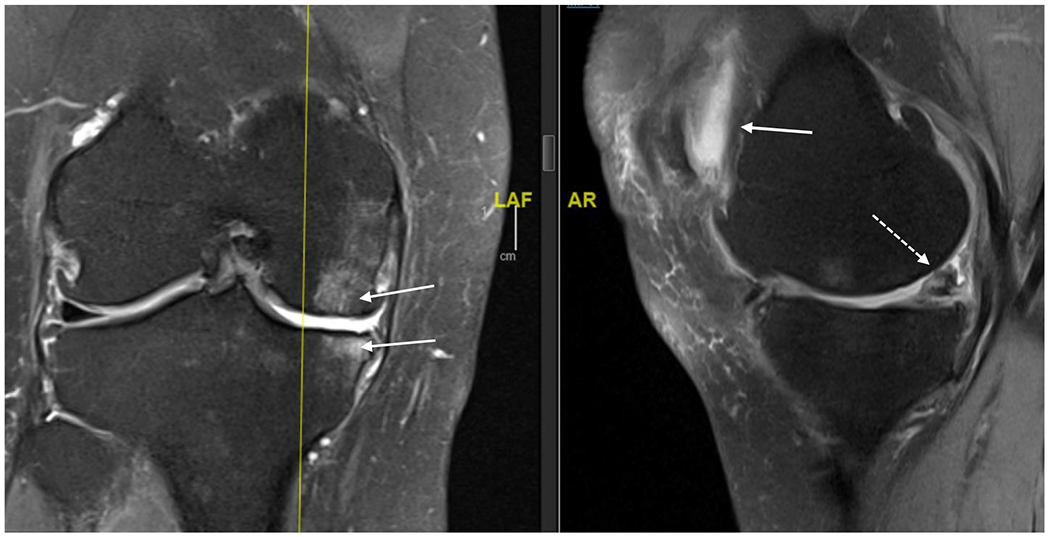
MRI (proton density, fat saturated) of right knee of 63 year old female. Coronal view on left and saggital view on right. Bone marrow lesions are identified with thin, solid white arrows on the coronal view; meniscal damage and cartilage damage are identified with dashed arrow on saggital view and retropatellar effusion as solid arrow on saggital view.