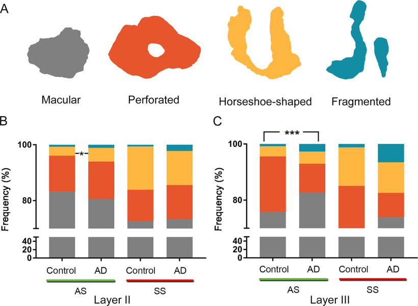
Figure 9.
Proportions of the different synaptic shapes (A) in Layers II (B) and III (C) of the EC in control subjects and AD cases. A, Schematic representation of the synaptic shapes: macular synapses, with a continuous disk-shaped PSD; perforated synapses, with holes in the PSD; horseshoe-shaped, with a tortuous horseshoe-shaped perimeter with an indentation; and fragmented synapses, with two PSDs with no connections between them. Proportions of macular, perforated, horseshoe-shaped, and fragmented AS and SS are displayed for Layers II (B) and III (C) of the EC in control subjects and AD cases. Statistical differences (indicated with asterisks) showed that in Layer II, horseshoe-shaped AS were more frequent (χ2, p = 0.04) in AD cases; and in Layer III of the AD group, fragmented (χ2, p = 0.0004) and macular (χ2, p < 0.0001) AS were more frequent, whereas perforated AS were less frequent in AD cases than in control individuals (χ2, p <0.0001).

