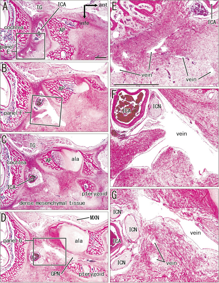
Fig. 4.
Wide and dense mesenchymal tissue containing the internal carotid artery and extending between the cochlear wall and sphenoid. Horizontal sections. A fetus of crown-rump length (CRL) 228 mm (approximately 28 weeks). H&E staining. (A) or (D) displays the most superior (or inferior) site in the figure. (E–G) are higher magnification views of squares in (A), (B), and (D), respectively. The left-hand side of each panel corresponds to the anterior side of the head. In these superior sections, no structure delineates the future carotid canal except for the bony alar process (AP) of the sphenoid. Instead, a dense mesenchymal tissue extends between the cochlear wall and sphenoid and it contains abundant veins. (A–D) or (E–G) were prepared at the same magnification; (A, E) Scale bar: 1 mm. ala, ala temporalis of the sphenoid; GPN, greater petrosal nerve; ICA, internal carotid artery; ICN, internal carotid nerve; MXN, maxillary nerve (intracranial course); TG, trigeminal ganglion.

