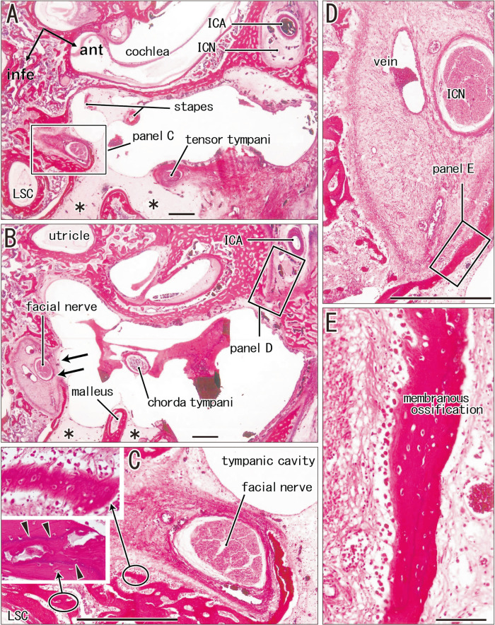
Fig. 7.
Carotid and facial nerve canals adjacent to the tympanic cavity: membranous ossification in these septa. Sagittal sections. A fetus of crown-rump length (CRL) 272 mm (approximately 31 weeks). H&E staining. The left-hand side of each panel corresponds to the posterior side of the head. Panel (A) displays a plane 1.2 mm medial to (B). (A, B) Contain the carotid and facial nerve canals: the septum is developing for the internal carotid artery (ICA), while the latter canal still opens widely to the tympanic cavity (arrows in B). (C) or (E) is a higher magnification view of a square in panel (A) or (D). The left-hand side of each panel corresponds to the anterior side of the head. Near the facial nerve, membranous ossification (upper insert of C) coexist endochondral ossification indicated by a tidemark (arrowheads; lower insert). (D) A section near (B), exhibits a site corresponding to a square in (B). (E) A higher magnification view of a square in (D), shows membranous ossification in a septum between the carotid canal and tympanic cavity. Scale bars: (A–D) 1 mm; (E) 0.1 mm. Magnification of inserts, same as (E). ant, anterior; infe, inferior; ICN, internal carotid nerve; LSC, lateral semicircular canal.

