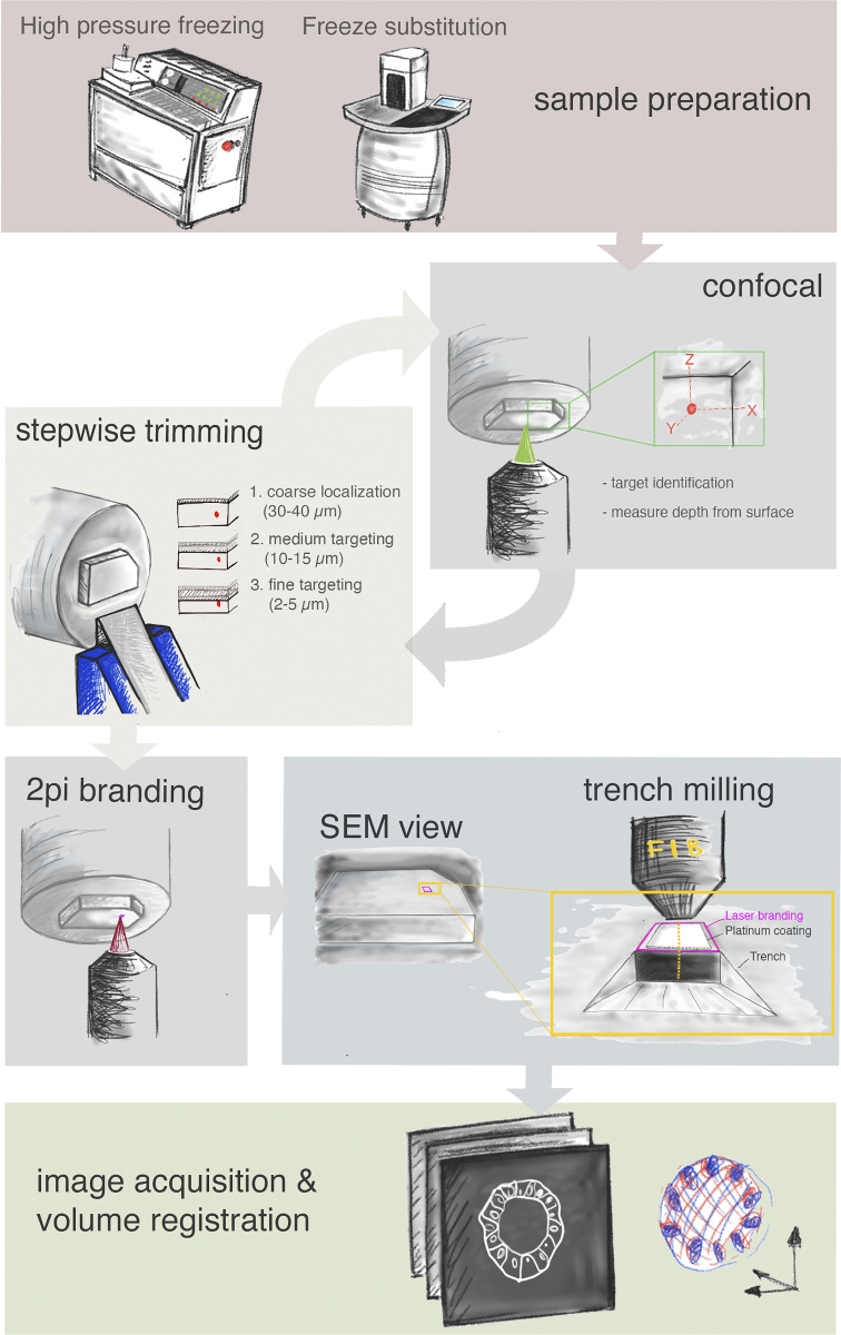
Figure 3.
Schematic representation of the workflow. From the top: Sample preparation consists of high-pressure freezing and FS. Second row: cycles of confocal acquisition and stepwise trimming at the ultramicrotome to progressively assess the depth of the target relative to the block surface. Normally, three iterations were sufficient, reaching each time the approximate distance in Z from target as indicated in the figure. Third row: The block surface is marked by two-photon (2Pi) branding (magenta). Preparation for FIB-SEM consists of placing the platinum coating and trench milling with the ion beam in the vicinity of the branded mark. Finally, FIB-SEM imaging and image processing in the last row include registration of the volumes obtained by FIB-SEM and confocal acquisition.

