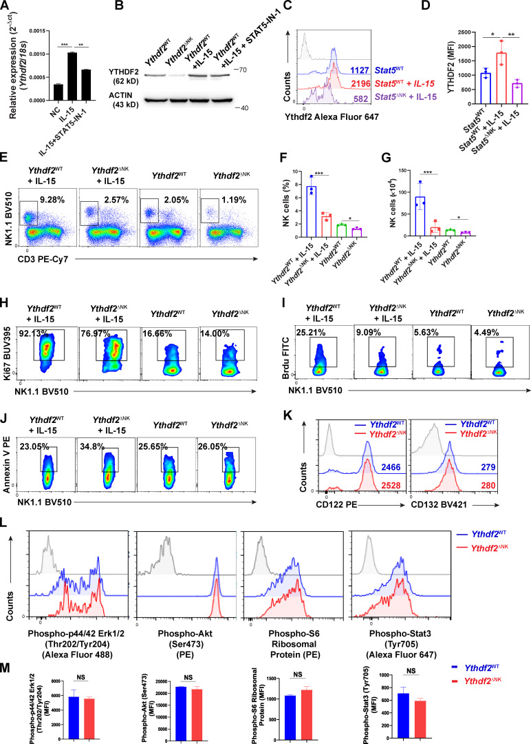
Figure S4.
IL-15-STAT5 signaling positively regulates YTHDF2 expression, and Ythdf2 deficiency does not affect the expression of IL-15 receptor components, the PI3K–AKT pathway, and the MEK–ERK pathway in NK cells.(A) qPCR showing the expression of Ythdf2 in NK cells at indicated time points following stimulation of IL-15. (B) qPCR showing the expression of Ythdf2 in NK cells in the presence of STAT5 inhibitor and IL-15 stimulation. (C and D) Representative histograms (C) and quantification (D) of intracellular staining showing the YTHDF2 protein levels in splenic NK cells from Stat5WT and Stat5ΔNK mice under the stimulation of IL-15. (E–G) Mice were treated with IL-15 (2 µg/d) for 5 d (n = 3 per group). The representative plots (E), percentages (F), and absolute numbers (G) of NK cells in the spleen are shown. (H–J) Representative plots of Ki67+ NK cells (H), BrdU+ NK cells (I), and annexin V+ NK cells (J) from IL-15–treated mice. (K) Representative histograms of CD122 and CD132 levels in splenic NK cells from Ythdf2WT and Ythdf2ΔNK mice. (L and M) Representative histograms (L) and quantification (M) of indicated molecules in splenic NK cells from Ythdf2WT and Ythdf2ΔNK mice after stimulation of IL-15. Data are shown as mean ± SD and were analyzed by unpaired two-tailed t test (M) or one-way ANOVA with Šídák post-test (A, D, F, and G). Data are representative of at least two independent experiments. *, P < 0.01; **, P < 0.01; ***, P < 0.001. MFI, median fluorescence intensity.

