Figure 1.
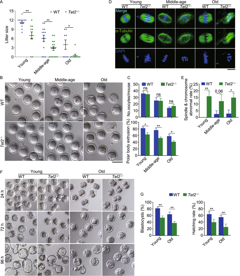
Tet2 knockout causes subfertility and impairs oocyte and early embryo development. (A) Average litter size of wild-type (WT) and Tet2 knockout (Tet2−/−) female mice at three different ages (young, 2–4 months; middle-age, 7–9 months; old, 10–12 months). n > 10 for successfully mated female mice for young and middle-age group and n ≥ 8 mice for old group are pooled. Each spot in round (blue) or square (green) shape represents the number of pups delivered from a successfully mated female mouse. (B) Representative images of superovulated oocytes from WT and Tet2−/− female mice at three different ages. Scale bar, 100 μm. (C) Comparison of oocyte yield per mouse (top) and percentage of the first polar body extrusion (bottom). n = 4–6 mice per group. (D) Representative images of oocytes stained with α-Tubulin antibody (green) and DAPI (blue) from three different age groups. Scale bar, 10 μm. (E) Spindle and chromosome abnormal rate between WT and Tet2−/− oocytes from three different age groups. n = 3–4 mice per group. (F) Morphology of embryo development at 2-cell (24 h), morula (72 h) and blastocyst (96 h) stage following in vitro fertilization of oocytes. Oocytes were obtained from WT and Tet2−/− mice from young or old group and sperm from healthy ICR male mice. (G) Percentage of developed blastocysts or blastocyst hatching rate 96 h after in vitro fertilization from young and old oocytes. n = 3–5 mice, and about 50 blastocysts were counted for each group. The bars indicate mean ± SEM. *P < 0.05; **P < 0.01; ns, no significant difference

