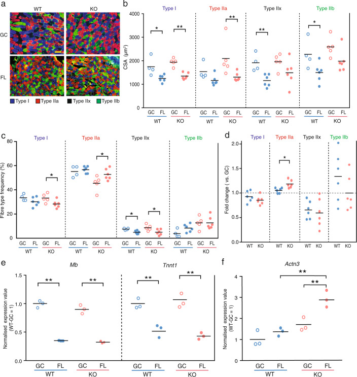
Fig. 5. Effect of Nrf2 deficiency on fibre type transition.
a Immunohistochemical staining for myosin heavy chain using BA-D5 (type I; blue), SC-71 (type IIa; red), and BF-F3 (type IIb; green) antibodies. Unstained fibres predicted to be type IIx (black). Scale bar = 100 μm. b CSA of each fibre type from WT-GC (n = 6), WT-FL (n = 6), KO-GC (n = 6) and KO-FL (n = 6). c Frequency of each fibre type in WT-GC (n = 6), WT-FL (n = 6), KO-GC (n = 6) and KO-FL (n = 6) mice. d Comparison of FL fibre type frequency normalised by GC fibre type frequency from WT and KO mice. P values from Student’s t tests (b–d) are indicated as follows: *P < 0.05, **P < 0.01, GC vs. FL. n = 6 for WT-FL and KO-FL, n = 5 for KO-GC and n = 4 for WT-GC. e Expression of Mb and Tnnt1 in soleus muscles. f Expression value of Actn3 in soleus muscles. Expression values were normalised by Quantile (WT-GC = 1). n = 3 for WT-GC, WT-FL, KO-GC and KO-FL. edgeR test (e, f) **P < 0.01 (FDR-corrected).

