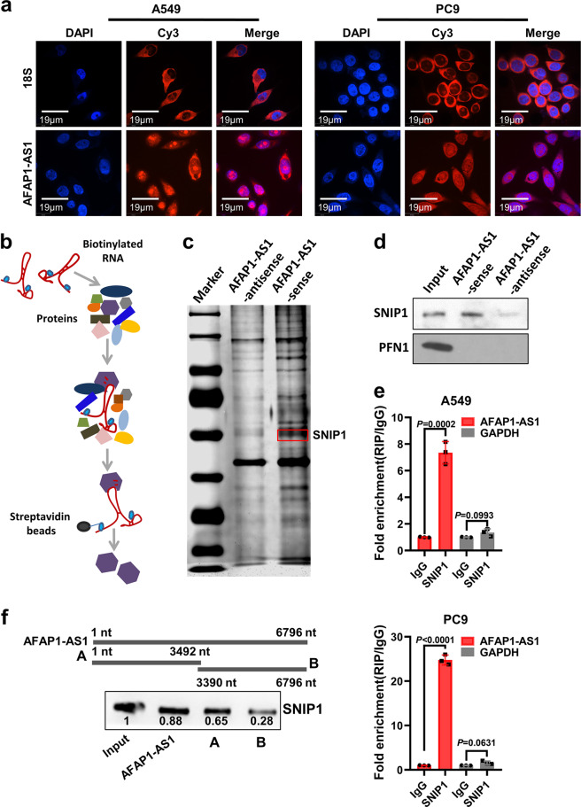
Fig. 3.
AFAP1-AS1 physically interacts with SNIP1 protein. a RNA FISH experiment was done to examine the localization and AFAP1-AS1 expression in A549 and PC9 cells. AFAP1-AS1 was distributed in both cytoplasm and nucleus. The RNA 18 S, localized in the cytoplasm, was used as a positive control. DAPI-stained nucleus: blue; Cy3 at the 5′ end of the probe (AFAP1-AS1 or 18 S): red; the merged image represents the overlap of DAPI and AFAP1-AS1 (Scale bar: 19 μm). b Schematic diagram of RNA pull down experiment performed to identify proteins of associated with AFAP1-AS1. Biotinylated sensor antisense or sense AFAP1-AS1 RNA was incubated with lysates of A549 cells targeted with streptavidin beads. Interacting proteins were resolved, visualized by silver staining, and identified by LC-MS/MS. c The result of silver staining of RNA pull down proteins. The red square indicates the approximate position of SNIP1. d Western blotting analysis shows that AFAP1-AS1 specifically interacts with SNIP1 protein but not PFN1 protein (a negative control). e RIP of AFAP1-AS1 in A549 and PC9 cells using anti-SNIP1 or IgG antibodies. The fold enrichment of AFAP1-AS1 is shown relative to that of the matched control IgG. RIP, RNA immunoprecipitation. f The deletion-mapping assay showed that the A fragment (1–3492 nt) of AFAP1-AS1 interacted with SNIP1 protein. Data are presented as the means ± SD

