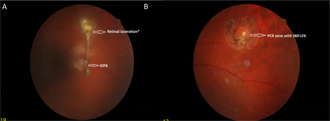
Fig. 5. Preoperative and postoperative retinal images of a patient with IOFB.
a The baseline fundus images of a patient with IOFB and retinal laceration. *The retina and choroid are damaged, and fibroblastic activity is seen. b The fundus image of the same patient following PPV, IOFB removal, and PCR with three rows of endolaser surrounding the zone.

