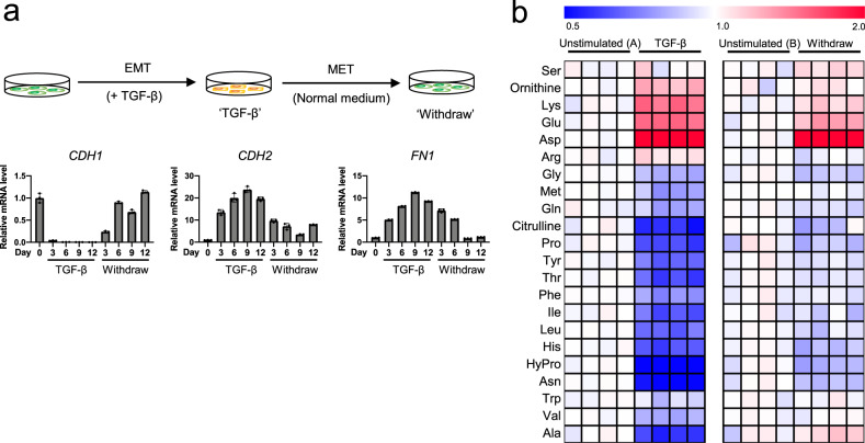
Fig. 2. Reversible EMT responses induced by TGF-β.
a Reversible EMT marker changes induced by TGF-β in A549 cells. A549 cells were stimulated with TGF-β (2 ng/mL) for the indicated number of days (marked TGF-β). After 12 days, the cells were washed with PBS and subsequently cultured in normal growth medium without TGF-β for 3, 6, or 9 days (marked Withdraw). The cells were passaged for 3 days. mRNA levels of EMT markers CDH1, CDH2, and FN1 were measured by real-time PCR. Values are presented as mean ± SD from triplicate samples. b TGF-β-induced reversible changes in amino acid levels in A549 cells. ‘TGF-β’, A549 cells stimulated with TGF-β (5 ng/mL) for 3 days, washed with PBS and subsequently cultured in regular medium without TGF-β for 3 days (‘Withdraw’). Controls were cultured in normal growth medium for 3 days (‘Unstimulated (A)’) and 6 days (‘Unstimulated (B)’) for TGF-β stimulation and withdrawal, respectively. The medium for ‘Unstimulated (B)’ was changed to fresh normal medium after culturing for 3 days, as well as for ‘withdraw’ cultures. Metabolite levels in each sample were converted to a fold-change relative to the average metabolite level of the paired non-stimulation. Red and blue indicate higher and lower levels, respectively, of metabolites in TGF-β-stimulated cells compared to those in the unstimulated cells (white).

