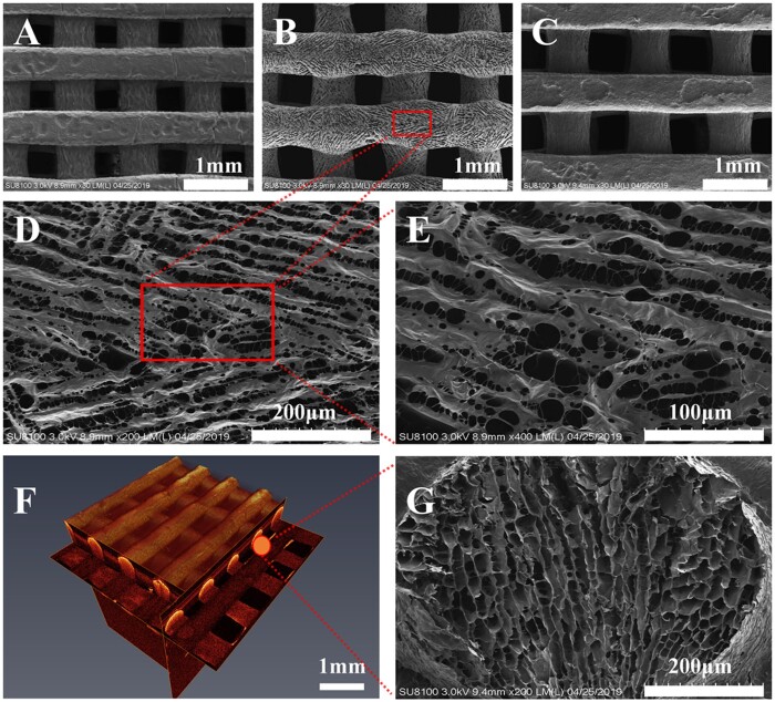
Figure 3.
The structure of the type I collagen/silk fibroin (CSF) scaffold. A–C: Scanning electron microscopy of scaffold materials with different aperture sizes (due to shooting reasons, A and C showed the bottom surface of scaffold materials, because the printing bottom plate was relatively straight under contact; B showed the top surface of scaffold materials, which the small pores on the surface of scaffold materials were clearly observed); B, D, E: Scanning electron microscopy observations of the CSF2 (magnification: ×200, ×400 and ×800, respectively); F: Optical coherence tomography (observations on the shape rule of scaffold material and uniform distribution of pore diameter); G: Cross-section of CSF2 (×200).

