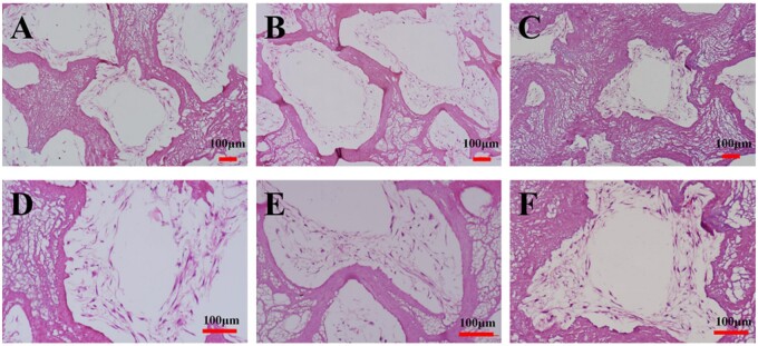
Figure 8.
HE staining of the HDPCs–CSF scaffolds cultured for 14 days. A, D: CSF1 group. HDPCs attached and spreaded within and around the scaffolds aperture; B, E: CSF2 group. HDPCs attached and spreaded similiar as in CSF1 group, formed multilayers; C, F: CSF3 group. Deformed structures of the scaffolds were observed in the CSF3 group.

